Anatomy Mapping Foramina - Foramen Magnum Hypoglossal Canal Jugular Foramen Anatomy Superior View Of The Skull Www Anatomynote Com Skull Anatomy Anatomy Bones Dental Anatomy / Foramen lacerum is an irregular opening located in the middle cranial fossa at the base of the skull.
Anatomy Mapping Foramina - Foramen Magnum Hypoglossal Canal Jugular Foramen Anatomy Superior View Of The Skull Www Anatomynote Com Skull Anatomy Anatomy Bones Dental Anatomy / Foramen lacerum is an irregular opening located in the middle cranial fossa at the base of the skull.. Foramen ovale is a foramen in the greater wing of sphenoid bone, and it gets its name from the latin word ovale, which means oval window. Endoscopic approach with bony landmarks. The lateral ventricles connected to the third ventricle by the interventricular foramina. Shahab shahid mbbs • reviewer: Learn vocabulary, terms and more with flashcards, games and other study tools.
This article discusses each of the aforementioned fossae and their associated foramina. In this article we will discuss the anatomy, its contents and clinical relevance of the mandibular mandibular foramen. The lesion originates at the left neural foramina and grows along the course of the brachial plexus (red arrow). The journal of laryngology clinical anatomy of blockade of the pterygopalatine ganglion: Uruj zehra mbbs, mphil, phd last.
2 from The optic foramen, the opening through which the optic nerve runs back into the brain and the large ophthalmic artery enters the orbit, is at the nasal side of the apex. Thoracic nerve roots emerge from the intervertebral foramina into the paravertebral space. (anatomy) an opening, an orifice, or a short passage, especially in a bone. View, isolate, and learn human anatomy structures with zygote body. It contains the incisive canals, which transmit palatine vessels and nerves. The lateral ventricles connected to the third ventricle by the interventricular foramina. Omental foramen or epiploic foramen or foramen of winslow or additus to lesser sac 【at t5 level】 (note: The journal of laryngology clinical anatomy of blockade of the pterygopalatine ganglion:
Frank pameijer, erik beek, frank joosten and robin smithuis.
Literature review and pictorial tour. The interventricular foramen, also known as foramen of monro, is part of the ventricular system and the connection between the third ventricle and the lateral ventricle. It contains the incisive canals, which transmit palatine vessels and nerves. Learn vocabulary, terms and more with flashcards, games and other study tools. Foundational anatomy provides medical students with the necessary background in. See more ideas about anatomy, muscle anatomy, arteries. A classic painting/stained glass window of foramen spinosum: Anatomy of the foramina of the skull. Foramen lacerum is an irregular opening located in the middle cranial fossa at the base of the skull. Borrowed from latin forāmen (aperture or opening produced by boring). Foramen (plural foramina or foramens). This article discusses each of the aforementioned fossae and their associated foramina. Each fossa contains specific foramina, through which various anatomical structures pass through.
The lesion originates at the left neural foramina and grows along the course of the brachial plexus (red arrow). The incisive foramen (foramen incisivum) is situated at the osseous palate behind the incisor teeth. Here they send white rami communicantes to the sympathetic chain. It is covered by cartilage after birth. Shahab shahid mbbs • reviewer:
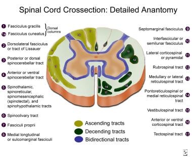
Topographic And Functional Anatomy Of The Spinal Cord Gross Anatomy Ventral And Dorsal Roots Descending Spinal Cord Tracts from img.medscapestatic.com Foundational anatomy provides medical students with the necessary background in. Greater petrosal nerve, deep petrosal nerve, internal carotid artery (all across upper portion). It is covered by cartilage after birth. Borrowed from latin forāmen (aperture or opening produced by boring). Thoracic nerve roots emerge from the intervertebral foramina into the paravertebral space. Applied knowledge of anatomical relations regarding surgical procedures and approaches was discussed and their relations were demonstrated via cadaveric dissections. The clinical impact of missed anatomy may result in failure and the necessity to carry out costly root the clinician must be aware of the complexities of the root canal system and anatomical variations of. Literature review and pictorial tour.
Shahab shahid mbbs • reviewer:
Foramen lacerum is an irregular opening located in the middle cranial fossa at the base of the skull. The incisive foramen (foramen incisivum) is situated at the osseous palate behind the incisor teeth. The journal of laryngology clinical anatomy of blockade of the pterygopalatine ganglion: Foramen ovale is a foramen in the greater wing of sphenoid bone, and it gets its name from the latin word ovale, which means oval window. Foramen (plural foramina or foramens). Omental foramen or epiploic foramen or foramen of winslow or additus to lesser sac 【at t5 level】 (note: Greater petrosal nerve, deep petrosal nerve, internal carotid artery (all across upper portion). Borrowed from latin forāmen (aperture or opening produced by boring). Here they send white rami communicantes to the sympathetic chain. Shahab shahid mbbs • reviewer: The clinical impact of missed anatomy may result in failure and the necessity to carry out costly root the clinician must be aware of the complexities of the root canal system and anatomical variations of. Uruj zehra mbbs, mphil, phd last. Lesser palatine nerve and vessels.
Each fossa contains specific foramina, through which various anatomical structures pass through. Shahab shahid mbbs • reviewer: Foramen (plural foramina or foramens). Greater petrosal nerve, deep petrosal nerve, internal carotid artery (all across upper portion). Borrowed from latin forāmen (aperture or opening produced by boring).
The Skull Anatomy And Physiology I from s3-us-west-2.amazonaws.com And sympathetic plexus $u!ular foramen inferior petrosal sinus lossopharyngeal, vagus and. View, isolate, and learn human anatomy structures with zygote body. It contains the incisive canals, which transmit palatine vessels and nerves. Anatomy of the foramina of the skull. This article discusses each of the aforementioned fossae and their associated foramina. A classic painting/stained glass window of foramen spinosum: Post ethmoidal foramen ant ethmoidal foramen frontoethmoidal suture anterior lacrimal crest optic foramen. In this article we will discuss the anatomy, its contents and clinical relevance of the mandibular mandibular foramen.
Location on base of skull foramen spinosum is adjacent to the spine of sphenoid.
Greater petrosal nerve, deep petrosal nerve, internal carotid artery (all across upper portion). Anatomy of the foramina of the skull. In the brain, the interventricular foramina (or foramina of monro) are channels that connect the paired lateral ventricles with the third ventricle at the midline of the brain. It is covered by cartilage after birth. Foramen ovale is a foramen in the greater wing of sphenoid bone, and it gets its name from the latin word ovale, which means oval window. A classic painting/stained glass window of foramen spinosum: As channels, they allow cerebrospinal fluid (csf). The lateral ventricles connected to the third ventricle by the interventricular foramina. The incisive foramen (foramen incisivum) is situated at the osseous palate behind the incisor teeth. Location on base of skull foramen spinosum is adjacent to the spine of sphenoid. Applied knowledge of anatomical relations regarding surgical procedures and approaches was discussed and their relations were demonstrated via cadaveric dissections. Post ethmoidal foramen ant ethmoidal foramen frontoethmoidal suture anterior lacrimal crest optic foramen. (anatomy) an opening, an orifice, or a short passage, especially in a bone.
As channels, they allow cerebrospinal fluid (csf) anatomy map. Here they send white rami communicantes to the sympathetic chain.


0 Komentar